 ultimate-guide
How to Read a Lumbar Spine MRI: A 2026 Guide
Table of Contents
- What Your Lumbar Spine MRI Actually Shows
- Understanding MRI Report Terminology: A Plain-Language Glossary
- Lumbar Disc Herniation Symptoms and What MRI Reveals
- AI-Powered MRI Report Analysis: How It Helps Patients
Last Updated: August 17, 2026
What Your Lumbar Spine MRI Actually Shows
A lumbar spine MRI is one of the most detailed diagnostic tools available for evaluating back pain, yet most patients receive results without real explanation. At Online Spine, we work with patients whose MRI reports are full of terminology that reads like a foreign language.
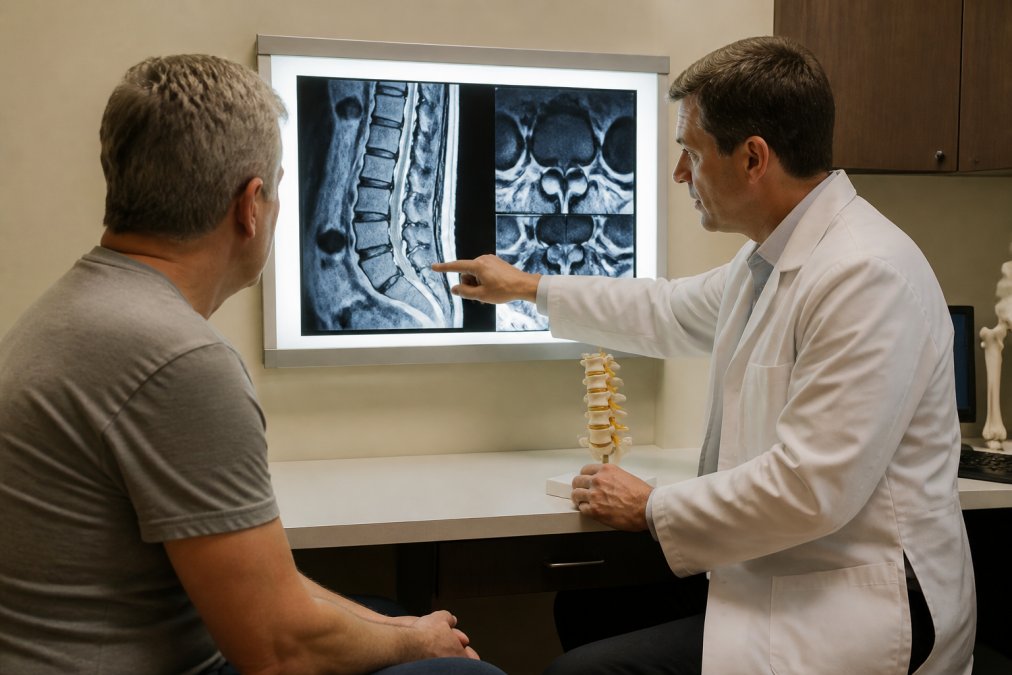
A lumbar spine MRI uses magnetic fields to produce detailed cross-sectional pictures of the five vertebrae in your lower back, along with the discs, nerves, and surrounding soft tissue. The scan captures images in multiple planes: the sagittal plane shows a side view of your spine, while the axial view gives a cross-section. Together, these views let a radiologist assess disc height, nerve root compression, and related structures.
Two image types dominate every lumbar spine MRI: T1-weighted and T2-weighted sequences. On T1-weighted images, fat appears bright and fluid appears dark. On T2-weighted images, fluid appears bright, making it ideal for spotting inflammation, disc degeneration, or nerve root impingement. Healthy, hydrated discs glow white on T2. Degenerated discs appear darker, indicating loss of water content.
Understanding MRI Report Terminology: A Plain-Language Glossary
Most guides skip the part that matters most to patients: what the words in the report actually mean. Here is a direct translation of the terms you are most likely to encounter.
Disc herniation is when the soft inner material of an intervertebral disc pushes through a tear in its outer ring, pressing toward nearby nerves. Herniation exists on a spectrum: disc protrusion (bulge with intact outer layer), disc extrusion (inner material breaks through outer layer), and sequestered fragment (piece breaks free and migrates into the spinal canal).
Spinal stenosis is narrowing of the spinal canal or the openings where nerve roots exit. Foraminal narrowing refers specifically to compression at those exit points, which can cause radiating pain, numbness, or tingling down the leg.
Signal intensity describes how bright or dark a structure appears on MRI. Hyperintense appears bright; hypointense appears dark. Modic changes are signal changes in the vertebral body adjacent to a disc, graded Type I through III, with Type I suggesting active inflammation.
Annular tear refers to a crack in the tough outer ring of the disc. Osteophytes are bony spurs that grow along vertebral edges in response to disc degeneration. Spondylolisthesis is when one vertebral body slips forward over the one below it. Facet joint hypertrophy is enlargement of the small joints at the back of each vertebra, often from arthritis.
According to the National Institute of Neurological Disorders and Stroke, many imaging findings like disc bulges and osteophytes are present in adults without any pain at all, a critical point that changes how you should read your own report.
| Term | Plain-Language Meaning | Common Symptom Link |
|---|---|---|
| Disc protrusion | Disc bulge, outer ring intact | Local back pain, possible nerve irritation |
| Disc extrusion | Disc material breaks through outer ring | Radiating leg pain, sciatica |
| Foraminal narrowing | Nerve exit point compressed | Shooting pain, numbness in leg or foot |
| Spinal stenosis | Spinal canal narrowed | Pain with walking, relief when seated |
| Modic changes | Bone marrow changes near disc | Chronic low back pain, inflammation |
| Spondylolisthesis | Vertebra slipped forward | Instability, nerve compression symptoms |
Lumbar Disc Herniation Symptoms and What MRI Reveals
The biggest disconnect between lumbar disc herniation symptoms and MRI findings is this: the severity of what shows up on the scan does not always match the severity of what the patient feels.
Lumbar disc herniation symptoms typically include sharp or burning pain traveling from the lower back down through the buttock and into one leg, a pattern called radiculopathy or sciatica. Numbness, tingling, or weakness in the foot or calf are also common. On MRI, the radiologist describes the herniation's location: central, paracentral, foraminal, or extraforaminal. A paracentral disc extrusion at L4-L5 can compress the L5 nerve root, producing symptoms along the outer shin and top of the foot.
The report may note nerve root impingement or thecal sac compression, both indicating the herniation is physically contacting neural structures. What the MRI cannot tell you is how much pain that contact is generating. The American Academy of Orthopaedic Surgeons clinical guidelines emphasize clinical correlation as the standard for interpreting spinal imaging findings.
What if the MRI shows no herniation but I still have leg pain?
Other findings can produce identical symptoms. Facet joint hypertrophy causing foraminal narrowing, ligamentum flavum thickening, or even a small sequestered fragment can all generate radiating pain without an obvious disc herniation.
AI-Powered MRI Report Analysis: How It Helps Patients
The gap between receiving an MRI report and understanding what it means has historically required a follow-up appointment, sometimes weeks away. AI-powered analysis is changing that timeline.

Online Spine provides an AI-guided, doctor-informed platform trained on over 500,000 real spinal cases. The system analyzes your MRI report and symptoms together, then delivers a plain-language explanation of your findings and a personalized recovery roadmap within minutes. Critical cases are flagged for manual review by verified spine specialists within 24 hours. The platform is fully HIPAA-compliant with end-to-end encryption protecting your data.
Research from the National Library of Medicine on AI in radiology shows AI-assisted analysis tools are increasingly effective at identifying clinically significant findings and supporting patient comprehension of complex imaging results.
Three questions worth asking your doctor after reviewing your MRI:
- Which specific finding on this MRI is most likely causing my current symptoms?
- Are any of these findings considered urgent, or is watchful waiting appropriate?
- What would need to change on a follow-up MRI to indicate I am improving or worsening?
Many patients experience significant anxiety after reading phrases like "moderate disc extrusion" or "severe foraminal narrowing" without context. A plain-language analysis that separates actionable findings from background noise is the difference between unnecessary panic and a clear path forward.
Frequently Asked Questions
How do I interpret a lumbar spine MRI report on my own?
Start by reading the 'Impression' or 'Conclusion' section at the bottom of the radiologist report, that is the summary of the most significant findings. Then work backward through the body of the report, which describes each spinal level. Look for terms like disc protrusion, foraminal narrowing, or nerve root impingement. A plain-language platform like Online Spine can translate these findings into clear, actionable language without requiring a medical background.
What does an abnormal lumbar spine MRI finding actually mean?
An abnormal finding on a lumbar spine MRI means the radiologist identified something that differs from typical anatomy, such as disc herniation, spinal stenosis, degenerative disc disease, or spondylolisthesis. Critically, many abnormal findings cause no symptoms at all. Clinical correlation, meaning how the imaging aligns with your actual pain and symptoms, is what determines whether a finding is clinically significant. Always discuss results with a qualified spine specialist before drawing conclusions.
What do white spots or bright signal changes mean on a spine MRI?
Signal intensity on MRI depends on which sequence you are viewing. On T2-weighted images, bright (hyperintense) areas often indicate fluid, inflammation, or disc degeneration. White spots near vertebral bodies may represent Modic changes, which signal bone marrow changes related to disc disease. On T1-weighted images, a bright signal can indicate fat. A radiologist report will specify which sequence shows the change and what it likely represents clinically.
What do terms like 'thecal sac' and 'foraminal stenosis' mean?
The thecal sac (also called the dural sac) is the membrane that surrounds the spinal cord and cauda equina. When a disc herniation or osteophyte presses against it, the report notes 'thecal sac compression.' Foraminal stenosis means narrowing of the bony opening (foramen) where nerve roots exit the spine, which can cause radiating pain, numbness, or tingling down the leg. Both terms describe how structural changes in the spine may be affecting nearby nerves.
Can I use an older MRI report if my pain has changed?
An older lumbar spine MRI can still provide useful structural context, but it may not reflect your current condition if symptoms have changed significantly. Disc herniations can resolve, worsen, or shift over time. A platform like Online Spine can analyze an existing report alongside your current symptoms to identify gaps and flag whether updated imaging is likely warranted, helping you decide your next step without immediately committing to a new scan.
How does AI-powered MRI report analysis differ from seeing a radiologist?
A radiologist reads the raw imaging and writes a technical report describing structural findings. AI-powered analysis, like the service offered by Online Spine, takes that report and translates it into plain language, correlates findings with your reported symptoms, and generates a personalized recovery roadmap. It does not replace a radiologist or treating physician, but it bridges the gap between a technical report and the actionable guidance most patients never receive after leaving the imaging center.
Understanding a lumbar spine MRI report is genuinely difficult without a clinical background. The terminology is dense, the findings are often multiple, and the connection between what the scan shows and what you actually feel is rarely straightforward. Online Spine bridges that gap with AI-guided analysis verified by spine specialists, delivering clear next steps without the wait. Start your spine assessment and get a personalized recovery roadmap based on your actual imaging findings.
This article was written using GrandRanker