 how-to
Doctor Verified Spine Recovery Plan: 2026 Guide
Table of Contents
- What a Doctor Verified Spine Recovery Plan Actually Includes
- Spine Surgery Recovery Timeline: What to Expect at Each Stage
- Evidence-Based Spine Rehabilitation Exercises for Functional Restoration
- How to Interpret Spinal Imaging Reports and Build Your Spine Recovery Plan
Last Updated: August 18, 2026
What a Doctor Verified Spine Recovery Plan Actually Includes
A doctor verified spine recovery plan is a structured, individualized roadmap combining clinical diagnosis, imaging analysis, and evidence-based treatment protocols to guide patients from acute pain toward functional restoration. A genuine plan integrates:
- Diagnosis clarification: Plain-language interpretation of MRI, CT, or X-ray findings, identifying disc herniation, spinal stenosis, spondylolisthesis, or nerve compression
- Pain management strategy: A tiered approach covering non-invasive options first, including anti-inflammatory protocols, activity modification, and targeted physical therapy
- Rehabilitation milestones: Specific, measurable goals at defined intervals (weeks 2, 6, 12, and 26 post-onset or post-surgery)
- Restrictions and precautions: Clear guidance on heavy lifting restrictions, bending mechanics, and posture correction to prevent re-injury
- Escalation criteria: Defined red flags that signal when conservative care should give way to orthopedic or surgical consultation
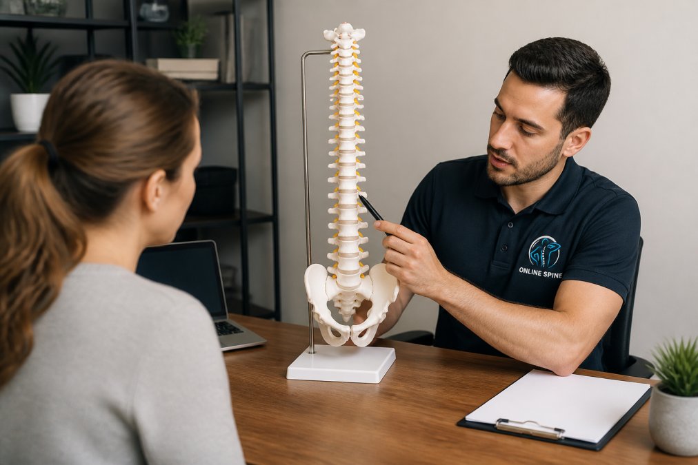
The difference between a generic plan and a personalized care plan comes down to specificity. A plan that doesn't account for your imaging findings, pain pattern, and functional limitations is, at best, a starting point.

Spine Surgery Recovery Timeline: What to Expect at Each Stage
Recovery after spinal procedures varies significantly depending on procedure type, from minimally invasive discectomy or laminectomy to multi-level spinal fusion. Most patients move through recognizable phases.
According to the American Academy of Orthopaedic Surgeons patient recovery guidelines, post-operative care for spinal procedures is divided into distinct recovery windows, each with different goals and restrictions.
Phase 1: Weeks 1-2 (Acute Recovery) The priority is swelling reduction and pain management. Most patients are restricted from bending, twisting, or heavy lifting. Walking short distances is encouraged to promote circulation and prevent blood clots.
Phase 2: Weeks 3-6 (Early Rehabilitation) Physical therapy typically begins, focusing on gentle range of motion and core activation. Patients recovering from laminectomy often progress faster than those post-fusion, where bone healing is the limiting factor.
Phase 3: Weeks 7-12 (Functional Restoration) Most patients return to light daily activities. The focus shifts to posture correction, mobility exercises, and progressive strengthening. Heavy lifting restrictions generally remain in place for fusion patients.
Phase 4: Months 3-6 (Return to Full Activity) Full functional restoration is the goal. Patients who engage consistently with rehabilitation and clinical oversight reach this milestone more reliably.
| Recovery Phase | Timeframe | Primary Goal | Key Restriction |
|---|---|---|---|
| Acute Recovery | Weeks 1-2 | Pain control, swelling reduction | No bending or twisting |
| Early Rehabilitation | Weeks 3-6 | Range of motion, core activation | Limit prolonged sitting |
| Functional Restoration | Weeks 7-12 | Strength, posture correction | Avoid heavy lifting |
| Return to Activity | Months 3-6 | Full mobility, work return | Cleared by surgeon |
Evidence-Based Spine Rehabilitation Exercises for Functional Restoration
The biggest mistake in spine rehabilitation is starting too aggressively. Functional restoration depends on a progressive, sequenced approach to mobility exercises.
The following exercises appear consistently across evidence-based rehabilitation programs. Always confirm with your treating clinician before beginning any exercise program.
Cat-Cow Stretch Improves spinal segmental mobility and reduces muscle tension along the thoracic and lumbar spine. Perform slowly, 10 repetitions per set, coordinating breath with movement.
Child's Pose Gently decompresses the lumbar spine and stretches the paraspinal muscles. Hold for 20-30 seconds. Patients with spinal stenosis may find this particularly relieving.
Pelvic Tilts and Bridge These activate the deep core stabilizers, essential for protecting spinal alignment during daily activities. Research from the National Institute of Neurological Disorders and Stroke on back pain rehabilitation supports core stabilization as foundational to neurological recovery and functional restoration.

How to Interpret Spinal Imaging Reports and Build Your Spine Recovery Plan
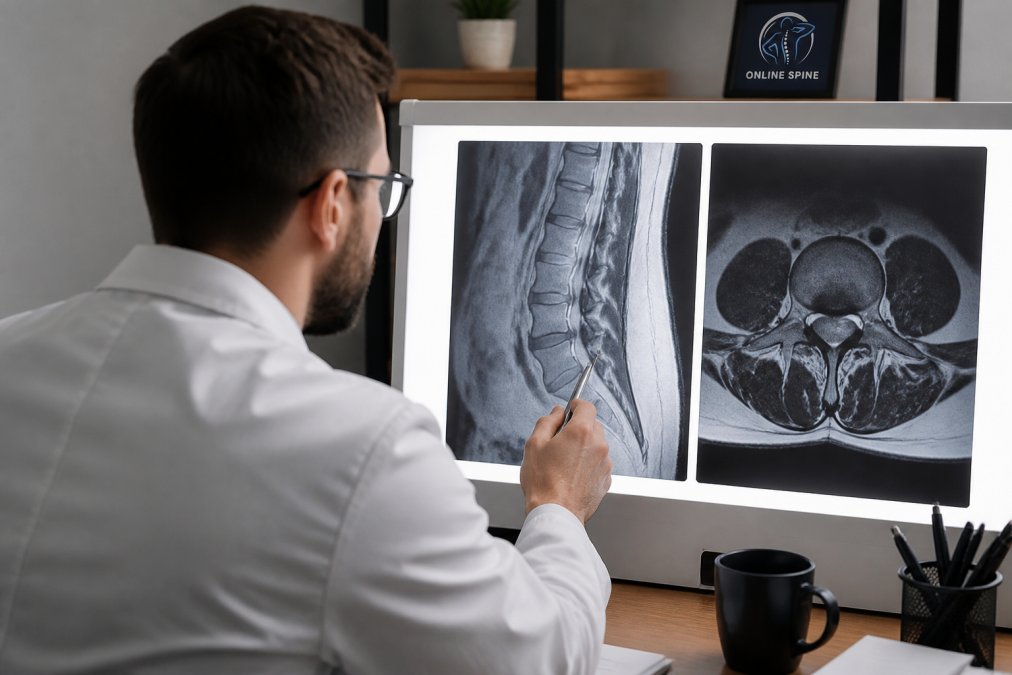
Spinal imaging reports are written for clinicians, not patients. Terms like "L4-L5 disc protrusion with mild neural foraminal narrowing" are accurate but nearly useless without context. Imaging findings only become useful when mapped to your symptoms and functional limitations.
Key terms to understand in your report:
- Herniation vs. protrusion vs. extrusion: These describe how far disc material has migrated. Extrusion is more severe than protrusion.
- Foraminal stenosis: Narrowing of the opening through which nerve roots exit the spine. A common cause of radiating pain and numbness.
- Modic changes: Signal changes in vertebral endplates on MRI, associated with discogenic pain.
- Spondylolisthesis grade: Describes how far one vertebra has slipped over another. Grade I is mild; Grade III-IV may require surgical evaluation.
According to the Radiological Society of North America's patient imaging resources, most spinal MRI findings need clinical correlation to be meaningful.
Building a spine recovery plan requires three steps: confirm which findings are symptomatic, match each symptomatic finding to a targeted intervention, and establish patient-reported outcome measures like the Oswestry Disability Index to track whether interventions are working over time.
A platform like Online Spine translates imaging findings into plain-language reports and maps them to actionable recovery roadmaps, verified by spine specialists. For patients managing radiating pain, numbness, or post-surgical recovery, that clarity is the difference between an effective spine recovery plan and months of guesswork.
A clear, personalized spine recovery plan requires accurate imaging interpretation, structured rehabilitation milestones, and consistent clinical oversight. If you're navigating a diagnosis alone or struggling to translate your MRI findings into a path forward, start with Online Spine's assessment process to receive a plain-language report and a doctor-verified roadmap built around your specific condition. Start Your Spine Assessment and stop guessing about what your spine actually needs.
Frequently Asked Questions
What are the key components of a doctor-verified spine recovery plan?
A clinically sound spine recovery plan typically covers four areas: an accurate diagnosis based on imaging and symptom history, a structured physical therapy and rehabilitation program, clear restrictions on activities like heavy lifting and bending, and a timeline of recovery milestones. The best plans also address pain management strategies, posture correction, and when to escalate care. Generic advice rarely accounts for whether your condition involves spinal fusion, laminectomy, discectomy, or conservative management, which is why personalized, doctor-verified guidance matters.
How long does spine surgery recovery take, and what affects the timeline?
Recovery after spinal surgery varies widely depending on the procedure. A microdiscectomy may allow a return to light activity within two to six weeks, while a multi-level spinal fusion can take six to twelve months for full functional restoration. Factors that influence the spine surgery recovery timeline include age, overall health, severity of nerve involvement, adherence to a rehabilitation program, and whether neurological recovery is complete. Your surgeon and physical therapist set the milestones, the plan should be revisited if pain or mobility changes significantly.
How do I know if my spine recovery plan is clinically sound?
A clinically sound plan is built on your specific imaging findings, not generic back pain advice. It should include measurable recovery milestones, a progression of mobility exercises, and clear guidance on restrictions like lifting limits. It should also specify when to seek urgent care, for example, if radiating pain, numbness, or weakness worsens. Platforms verified by licensed spine specialists and trained on large case datasets can help translate your MRI or X-ray findings into a personalized care plan, but they do not replace direct clinical oversight.
Can AI-powered tools give useful spine recovery guidance without a recent MRI?
Yes, with some caveats. Symptom patterns and prior imaging can still generate meaningful guidance even if your scan is several months old. However, if your pain has changed significantly, especially if you have new radiating pain, numbness, or weakness, updated imaging gives a more accurate picture of your current spinal condition. A good AI-assisted platform will flag cases where clinical review is needed and will be transparent about the limits of older data, rather than issuing a recovery roadmap that may no longer reflect your actual condition.
This article was written using GrandRanker